Hello, this is Goyang Shinsegae Eye Clinic.
After wearing glasses or contact lenses for many years, some people begin considering vision correction surgery because of the inconvenience of daily use and maintenance.
At this point, you may wonder not only how LASIK and LASEK differ, but also which equipment is used and which procedure may be suitable for your eye condition.
Vision correction surgery should be planned after carefully evaluating factors such as corneal thickness and shape, the degree of myopia and astigmatism, and overall eye health—not visual acuity alone.
AMARIS RED LASEK is characterized by its small laser beam, eye-tracking technology, and real-time corneal thickness measurement system.
Today, we will examine the differences between 2-Day LASEK and SMILE LASIK, the main features of AMARIS RED LASEK, and what you should consider before choosing a vision correction procedure.

SMILE LASIK vs. 2-Day LASEK
✔ Corneal Tissue Removal and Glare
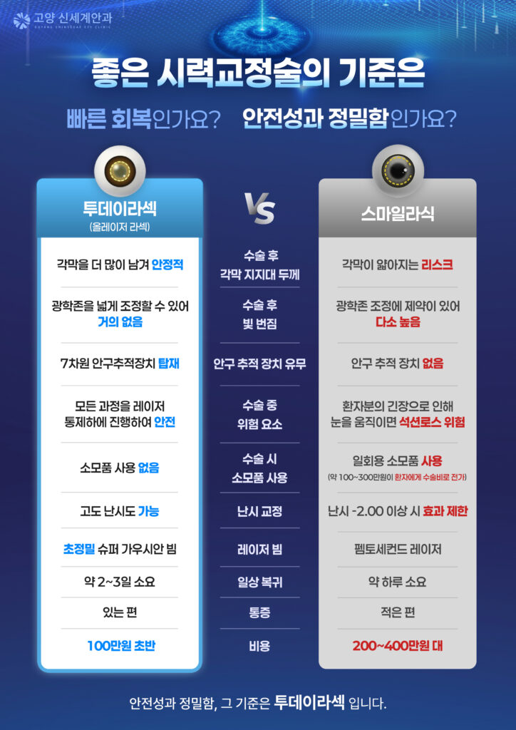
2-Day LASEK can preserve more of the cornea’s structural support, which may help reduce the risk of secondary complications such as corneal ectasia.
SMILE LASIK, on the other hand, may leave less structural support than LASEK, potentially resulting in a comparatively greater risk in some patients.
*Corneal ectasia: A complication in which a weakened area of the cornea gradually protrudes after LASIK or SMILE surgery because the remaining corneal tissue is insufficient.
✔ Intraoperative Stability and Treatment Decentration
LASIK, SMILE, and LASEK procedures require accurate treatment of an optical zone approximately 6 mm in diameter at the center of the cornea.
However, patients may make small eye movements during the procedure because of tension or nervousness, making accurate targeting difficult without a tracking system.
For this reason, excimer laser systems are equipped with eye-tracking technology. This system monitors eye movements and helps keep the laser aligned with the planned treatment area.
SMILE LASIK does not use the same active eye-tracking system during laser application. Instead, suction is used to hold the eye in place.
If suction is interrupted because of eye movement or tension, a condition known as “suction loss” may occur. Some reports have estimated that suction loss occurs in approximately four out of every 100 cases.
With 2-Day LASEK, the procedure is performed under laser control. SMILE LASIK, however, requires the surgeon to manually separate and remove the lenticule. This step may involve additional procedural risks, such as irregular tissue tears or epithelial ingrowth within the corneal stroma.
✅ Astigmatism Correction
2-Day LASEK may be considered for patients with high myopia or high astigmatism.
With SMILE LASIK, the predictability of astigmatism correction may become more limited when astigmatism exceeds −2.00 D.
✅ Glare and Halos
2-Day LASEK allows greater flexibility in adjusting the optical zone, which may help reduce postoperative glare and halos.
SMILE LASIK may offer less flexibility in optical-zone adjustment, so the likelihood of glare or halos may be comparatively higher in some patients.
✅ Cost of Surgery
2-Day LASEK generally costs slightly over KRW 1 million, whereas SMILE LASIK may cost between KRW 2 million and KRW 4 million.
One reason for this difference is that SMILE LASIK requires a disposable suction interface. Depending on the equipment, this single-use component may cost approximately KRW 1 million to KRW 3 million, which is reflected in the overall surgical fee.
Because 2-Day LASEK does not require similarly expensive disposable components, it may be a more economical option.
What Are the Features of AMARIS RED LASEK?
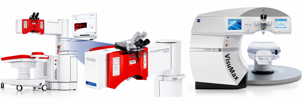
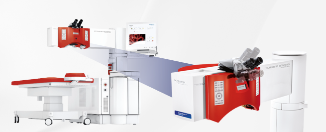
AMARIS RED is an excimer laser system used for LASEK and other laser vision correction procedures.
Its main features include a seven-dimensional eye-tracking system and a real-time corneal thickness measurement system.
Even when a patient focuses on a single point, the eye may still make subtle movements.
The seven-dimensional eye-tracking system detects movements in multiple directions during the procedure and helps deliver the laser to the planned treatment area.
The system can also monitor corneal thickness during the procedure, helping the surgeon assess whether treatment is progressing within the preoperatively planned range.
When considering AMARIS RED LASEK, it is advisable to look beyond the name of the equipment and understand how these features are used during the procedure.

It Uses a Small 0.54 mm Laser Beam
AMARIS RED uses a laser beam measuring 0.54 mm in diameter.
This small beam can be used to treat the intended area in greater detail.
Because the cornea is not completely flat, the laser must be applied while considering its shape and the required amount of correction in each area.
AMARIS RED LASEK uses a 0.54 mm laser beam to deliver detailed treatment across the planned area, with the goal of creating a smoother corneal surface.
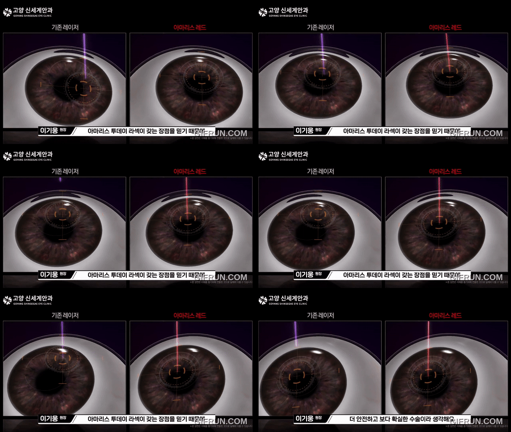
AMARIS RED LASEK Focuses on the Necessary Treatment Area
Conventional LASEK may use alcohol or a separate instrument to remove the corneal epithelium.
AMARIS RED LASEK differs in that a laser can also be used during the epithelial removal stage.
The epithelial removal area is reported to be approximately 63.6 mm² with conventional methods and 38.4 mm² with the AMARIS RED method. The procedure is designed to apply the laser primarily to the required optical area.
Reducing the use of alcohol or a brush for direct epithelial removal may also limit chemical and mechanical contact with the cornea.
However, the actual treatment area may vary depending on the patient’s refractive error and eye condition. A comprehensive examination is therefore necessary before surgery.

Even with the same AMARIS RED LASEK procedure, suitability may vary depending on the degree of myopia and astigmatism, corneal thickness and shape, pupil size, and the presence of dry eye disease.
Rather than choosing a procedure based solely on the features of a particular device, the first step should be a detailed examination to evaluate your current eye condition.
Please consult an ophthalmologist, confirm which method is appropriate for your eyes, and make your decision carefully.
Thank you.