

Quick answer: The key warning signs of retinal detachment are a sudden increase in floaters, flashes of light, and a dark shadow or “curtain” spreading across part of your vision. These symptoms need a dilated eye exam the same day, because early treatment gives the best chance of preserving sight.
Whether you live in Unjeong New Town, Geumchon, or central Paju, it can be tempting to wait and see whether odd visual symptoms settle overnight. With retinal detachment, waiting can matter. This guide explains what retinal detachment is, the symptoms that should never be ignored, who is at risk, and what to do if symptoms start in the evening or on a weekend.
What Is Retinal Detachment?
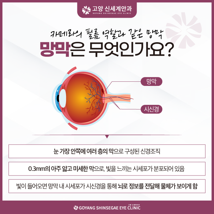
The retina is the thin, light-sensitive layer lining the back of the eye. It works like the sensor in a camera, converting light into signals that travel to the brain. Retinal detachment happens when the retina separates from the tissue beneath it, which supplies oxygen and nutrients. A detached retina cannot function properly, and if the detachment reaches the macula, the center of the retina responsible for fine detail, central vision can be seriously affected.
Three main types
- Rhegmatogenous detachment: the most common type. A tear or hole in the retina lets fluid from the vitreous gel pass underneath and lift the retina.
- Tractional detachment: scar tissue on the retinal surface pulls it away, often seen in advanced diabetic retinopathy.
- Exudative detachment: fluid builds up beneath the retina without a tear, due to inflammation, tumors, or vascular conditions.
Warning Signs of Retinal Detachment
Retinal detachment itself is usually painless, which is why people sometimes delay. Watch for:
- A sudden shower of new floaters: dots, cobwebs, or specks drifting across your vision, especially if many appear at once.
- Flashes of light (photopsia): brief sparks or lightning streaks, often at the edge of vision and more noticeable in dim light.
- A shadow or curtain: a gray or dark area spreading from the side, top, or bottom of your visual field.
- Blurred or distorted central vision: straight lines may look wavy if the macula is involved.
- Sudden decrease in vision in one eye.
Why the “curtain” matters most
Floaters and flashes can come from a harmless posterior vitreous detachment (PVD), but the same process can tear the retina. A spreading shadow suggests the retina may already be lifting. Any of these symptoms, especially when new, should be evaluated urgently rather than at a routine appointment.
Who Is at Higher Risk?
- High myopia: strongly nearsighted eyes are longer, and the retina is stretched thinner. This is particularly relevant in Korea, where myopia rates are high.
- Age over 50, when the vitreous gel naturally liquefies and separates.
- Previous cataract surgery or other eye surgery.
- Previous retinal tear or detachment in either eye.
- Family history of retinal detachment.
- Eye trauma, including sports injuries.
- Lattice degeneration, thin areas of peripheral retina that may be found on a dilated exam.
People who have had LASIK or LASEK should remember that laser vision correction changes the cornea, not the length of the eye. The retinal risk associated with high myopia remains after refractive surgery.
How Is Retinal Detachment Diagnosed?
Dilated fundus examination
After dilating drops, the ophthalmologist examines the entire retina, including the far periphery where most tears occur, often with indentation of the eyelid to view the edges.
Wide-field retinal imaging and OCT
Wide-field photographs document the peripheral retina, while OCT shows whether the macula is attached, which helps guide urgency and treatment planning.
Ultrasound
If bleeding inside the eye blocks the view, B-scan ultrasound can reveal whether the retina is detached.
How Are Retinal Tears and Detachments Treated?
Treatment depends on what is found:
- Retinal tear without detachment: often treated with laser photocoagulation or cryotherapy to create a seal around the tear. This is usually an in-office procedure.
- Retinal detachment: generally requires surgery, such as vitrectomy (removing the vitreous gel and flattening the retina, often with a gas or silicone oil bubble), scleral buckle, or pneumatic retinopexy, depending on the case.
After surgery with a gas bubble, specific head positioning may be required, and air travel or high altitude must be avoided until the gas absorbs. Visual recovery varies and depends heavily on whether the macula was involved and how long the detachment was present.
What Should Paju Residents Do If Symptoms Start?
- Do not wait to see if it goes away. Call an eye clinic the same day.
- During our clinic hours, contact us at 031-8076-9999 and describe your symptoms so we can advise how quickly to come in.
- Outside clinic hours (evenings, Saturday afternoons, Sundays), go to the emergency department of a hospital with an on-call ophthalmologist rather than waiting until the next weekday.
- Avoid strenuous activity, heavy lifting, and contact sports until you have been examined.
- Bring a list of medications and mention any previous eye surgery or high myopia.
Distance from your clinic should not be a reason to delay. If you cannot reach us quickly, the nearest emergency eye care is the right choice.
Can Retinal Detachment Be Prevented?
Not every case can be prevented, but risk can be reduced:
- Have a dilated retinal exam if you are highly myopic, before and after refractive surgery, and after cataract surgery as advised.
- Treat retinal tears found on screening promptly.
- Wear protective eyewear for sports and DIY work.
- Learn the warning signs and act on them quickly.
Frequently Asked Questions
Is retinal detachment painful?
Usually not. Retinal detachment is typically painless, which is why sudden floaters, flashes, or a shadow in vision should be taken seriously even without pain.
How quickly should I see a doctor for sudden floaters and flashes?
Ideally the same day. A dilated eye exam can determine whether there is a retinal tear or detachment, and tears found early can often be treated before a detachment develops.
Can a detached retina heal on its own?
A true retinal detachment does not usually reattach on its own and generally needs surgical repair. Small tears without detachment may be sealed with laser to prevent progression.
Does high myopia increase the risk of retinal detachment?
Yes. Highly nearsighted eyes are longer and have a thinner, more stretched retina, which raises the risk of tears and detachment. Regular dilated exams are recommended.
Will my vision return to normal after retinal detachment surgery?
It varies. Outcomes depend largely on whether the macula was detached and for how long. Earlier treatment generally offers a better chance of preserving vision, but no outcome can be guaranteed.
Key Takeaways
- Sudden floaters, flashes, and a spreading shadow are the classic warning signs of retinal detachment.
- Retinal detachment is usually painless, so absence of pain is not reassurance.
- High myopia, age over 50, prior eye surgery, and family history raise the risk.
- Same-day dilated examination is recommended; outside clinic hours, use hospital emergency eye care.
- Tears found early may be sealed with laser; detachments usually require surgery.
Medical disclaimer: This article is provided for general health information and education only. It is not a diagnosis or a substitute for an in-person examination. Suitability of any test or treatment, and its results, differ from person to person, and every procedure carries potential risks and side effects. Please consult an ophthalmologist about your own eyes. Regulatory approvals mentioned (such as US FDA decisions) apply only to the jurisdiction named, and the products discussed may not be approved or available in Korea.
Goyang Shinsegae Eye Clinic (고양신세계안과)
- Address: 6F Haengsin Square, 126 Chungjang-ro, Deogyang-gu, Goyang-si, Gyeonggi-do, Korea
- Phone: 031-8076-9999
- Hours: Mon–Fri 09:00–18:30 · Sat 09:00–14:00 · Lunch 13:00–14:00 · Closed Sundays